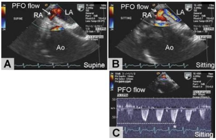
Figure 2.
Transesophageal echocardiography with color Doppler at the level position showing a patent foramen ovale (PFO) behind the enlarged aortic root (Ao). (A) In the supine position, paradoxical right-to-left shunt through the PFO is transiently observed as only a stream. (B) In the sitting position, the shunt grows to a massive jet. (C) A continuous-wave Doppler recording shows that shunt flow is observed in the systolic phase, with a peak velocity of 0.86 m/s (asterisk) and a calculated pressure gradient of 2.9 mmHg. (LA = left atrium; RA = right atrium.)

