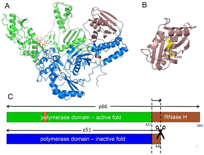
Figure 1.
Structure of the reverse transcriptase (RT) heterodimer. (A) Ribbon diagram of p66/p51 showing the active polymerase (green) and ribonuclease H (RNase H, brown) domains in p66, and the inactive polymerase (blue) and RNase H domain fragment, residues 427–440 (brown), in the p51 subunit. The YMDD motif at the active site in p66 is shown in orange; (B) Structure of the isolated RNase H domain identifying the buried Phe440–Tyr441 cleavage site (yellow); (C) Domain structure of the two RT subunits color coded as in A, and illustrating the internal cleavage site that produces the p51 subunit.

