Figure 4.
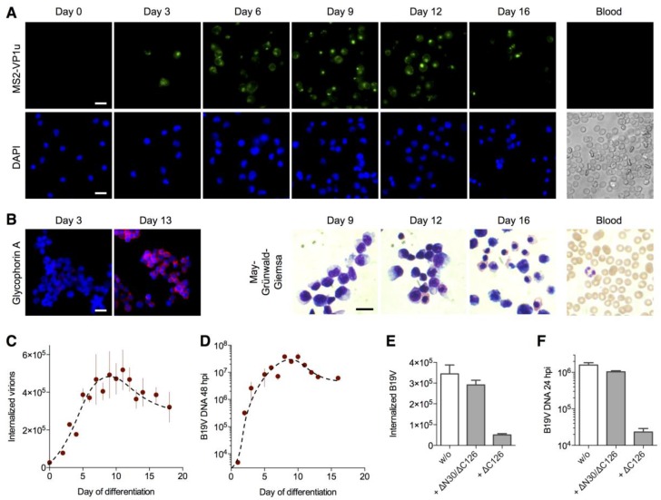
VP1u receptor expression correlates with B19V internalization and infection along the erythroid differentiation: (A) cluster of differentiation (CD) 34+ hematopoietic stem cells were expanded ex vivo to differentiate into the erythroid lineage and tested for MS2-∆C126 VP1u uptake (63× magnification). MS2-∆C126 VP1u internalization was further tested on peripheral blood cells, containing mature erythrocytes and ~1% reticulocytes, representing the final erythroid differentiation stages; (B) Immunostaining of glycophorin A (63× magnification) and May–Grünwald–Giemsa staining (100× magnification) indicates the successful transition to the terminal erythroid differentiation; B19V internalization (C) and infection (D) along the erythroid differentiation was quantified by qPCR (n = 3); (E) Internalization of B19V into erythroid progenitor/precursor cells (day 8) was competed with ∆C126 VP1u or dysfunctional ∆N30/∆C126 VP1u; (F) After VP1u-competed uptake of B19V, cells (day 8) were incubated for further 24 h to measure the effect of the competition on the viral infection (n = 3). Notably, B19V internalization was performed with more cells (2×) and higher multiplicity of infection (MOI) (7.5×) compared with infectivity assays, explaining the relatively high detection signal of uptake versus infection. Scale bar: 15 µm.

