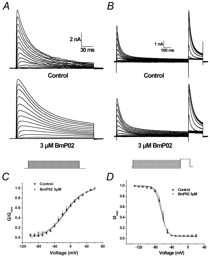
Figure 2.
Voltage-dependent activation and inactivation of Kv4.2 before and after application of 3 μM BmP02: (A) representative traces of K+ currents elicited by stimulus voltages from −100 mV to +60 mV in 10 mV increment from holding potential of −100 mV in control (top) and in the presence of 3 μM BmP02 (bottom); (B) voltage dependence of steady-state inactivation determined by a two-step protocol in which a conditioning pulse to potentials ranging from −130 mV to +20 mV was followed by a test pulse to +40 mV to measure the peak current amplitudes; and (C,D) normalized G-V relationship and I-V relationship in the absence (black squares) and presence (grey triangles) of 3 μM BmP02. The fitting parameters are indicated in Table 1. n = 10; mean ± S.E.M.

