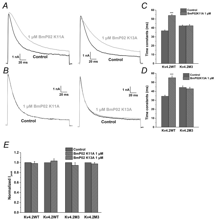
Figure 6.
Analysis of basic amino acid residues in BmP02: (A) Representative currents of Kv4.2 in the absence (black) and presence (grey) of the 1 μM BmP02 K11A (left) or BmP02 K13A (right). (B) Representative currents of Kv4.2M3 in the absence (black) and presence (grey) of the 1 μM BmP02 K11A (left) or BmP02 K13A (right). (C,D) Time constants of inactivation of Kv4.2 and Kv4.2M3 in the depolarization of +40 mV before (black square) and after (grey triangle) application of 1 μM BmP02 K11A or BmP02 K13A, respectively. n = 10; mean ± S.E.M. *** p < 0.001, significant difference between Control and BmP02 K11A or BmP02 K13A. (E) Normalized peak current of the Kv4.2 or Kv4.2M3 evoked by +40 mV pulses in the absence (black) or presence (grey) of 1 μM BmP02 K11A or BmP02 K13A.

