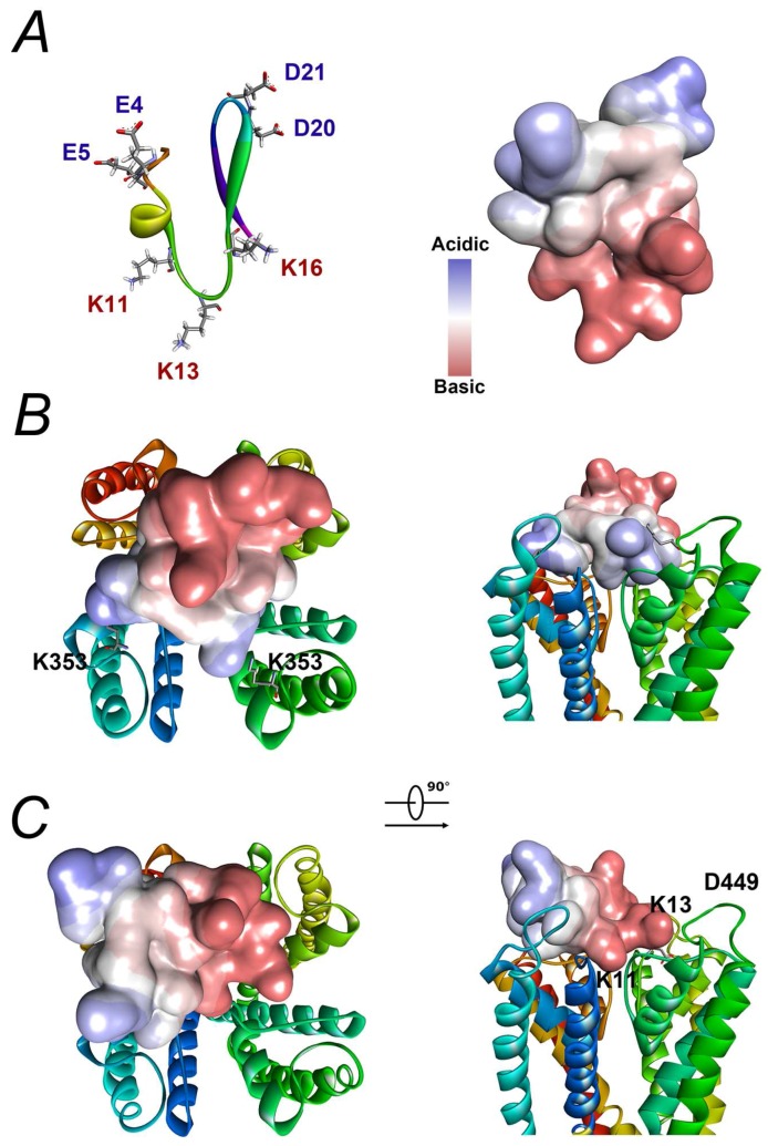
Figure 8.
Docking analysis of BmP02 on Kv4.2 and Kv1.3: (A) The three-dimensional structure of BmP02 the NMR study (PDB: 1DU9). Basic acids are highlighted in red, while acidic ones are in blue (left). A molecule surface is generated to show the acid-Base Properties of BmP02 (right). (B) The action mode of BmP02 on Kv4.2. The highlighted K353 in two subunits determines the action mode of BmP02 described in the text. (C) A model of BmP02 binding to Kv1.3 is a classical model with a lysine (K11) using its side chain plugging the channel. K13 in BmP02 and D449 in Kv1.3 are highlighted.

