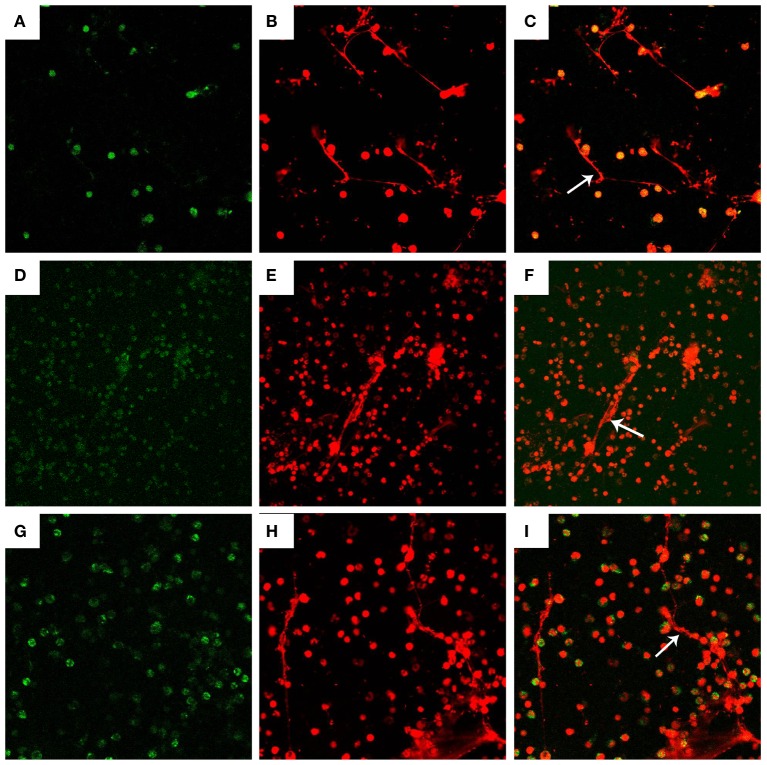
Figure 2.
Visualization of DNA decorated with histones (H3), neutrophil elastase (NE), and myeloperoxidase (MPO) in N. caninum tachyzoite-induced NETs structures. PMN were stimulated with Neospora caninum (ratio: 1:1, 90 min). DNA decorated with of H3, NE, and MPO within NETs were detected using a scanning confocal microscope. (A) Histone (Green). (D) MPO (Green). (G) NE (Green). (B,E,H) DNA (Red). (C,F,I) Respective merge of DNA decorated with H3, NE, and MPO.

