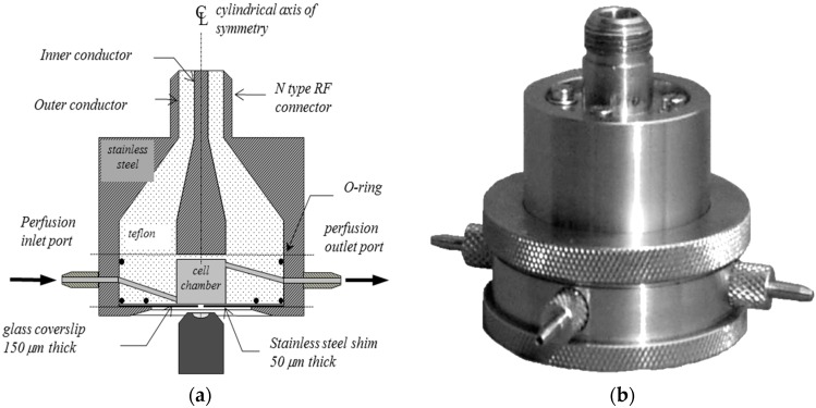
Figure 1.
The RF chamber consisting of a cell medium chamber at the end of a modified coaxial transmission line. It is designed to fit on any inverted microscope stage and is used with standard microscope objectives. A glass coverslip forms the floor of the cell chamber and the biological material is viewed through a central 1 mm diameter hole in an underlying steel shim via laser scanning confocal microscopy. The shim shields the cells from RF reflections from the objective. The chamber is shown in vertical cross-section in (a); and in perspective view in (b). Normally, a coaxial cable from the RF signal generator is attached at the top and the side ports are used to allow perfusion or temperature measurement via fluor-optic probes.

