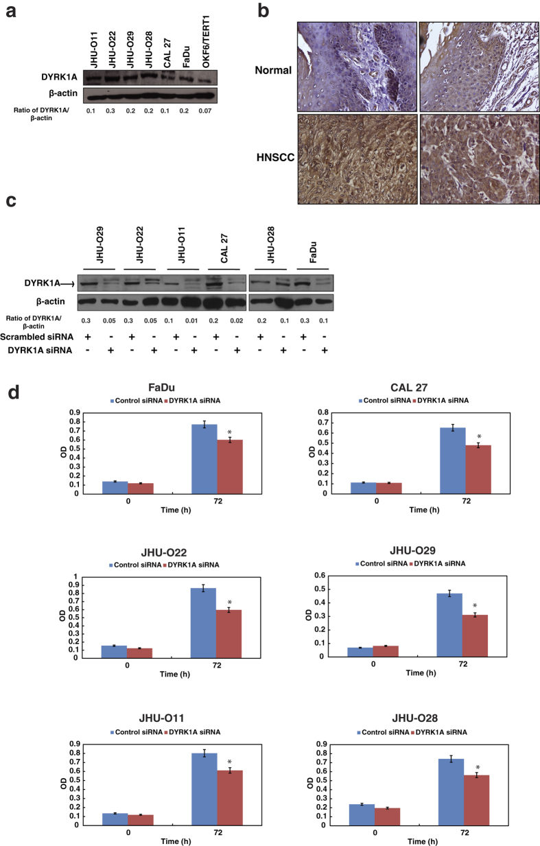
Figure 1. Inhibition of DYRK1A reduces cellular proliferation in HNSCC.
(a) Western blot analysis shows the expression profile of DYRK1A in a panel of HNSCC cell lines – JHU-O11, JHU-O22, JHU-O28, JHU-O29, FaDu and CAL 27 compared to normal oral keratinocytes OKF6/TERT1. (b) Immunohistochemical validation of DYRK1A in HNSCC tissue - representative sections from normal and HNSCC cases were stained with anti-DYRK1A antibody. (c) Western blot analysis depicting DYRK1A expression in HNSCC cell lines upon transfection with DYRK1A siRNA. β-actin was used as a loading control. (d) Cellular proliferation of HNSCC cells upon siRNA mediated silencing of DYRK1A (*p < 0.05).

