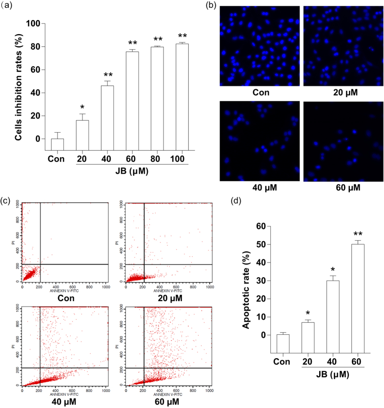
Figure 1. JB inhibits B16F10 cell growth and induces cell apoptosis.
(a) Cell viability was determined by SRB assay after 24 h treatment with different concentrations of JB (20, 40, 60, 80 and 100 μM). (b) Morphologic measurements in B16F10 cells were carried out via Hoechst fluorescence staining. (c) Representative images showing the apoptotic cells after treatment with the indicated concentrations of JB. (d) The apoptotic rates of B16F10 cells after JB treatment. The data represent mean ± s.d. of the three independent experiments. *P < 0.05, **P < 0.01 compared with the control group.

