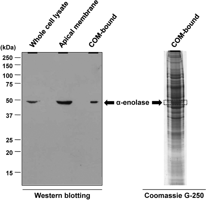
Figure 1. Western blot analysis of α-enolase.
Proteins in whole cell lysate, apical membrane and COM crystal-bound fractions were resolved by 12% SDS-PAGE and subjected to Western blot analysis using rabbit polyclonal anti-α-enolase (Santa Cruz Biotechnology) as a primary antibody. Coomassie Brilliant Blue G-250-stained gel of the COM-bound fraction was also aligned with the immunoblot.

