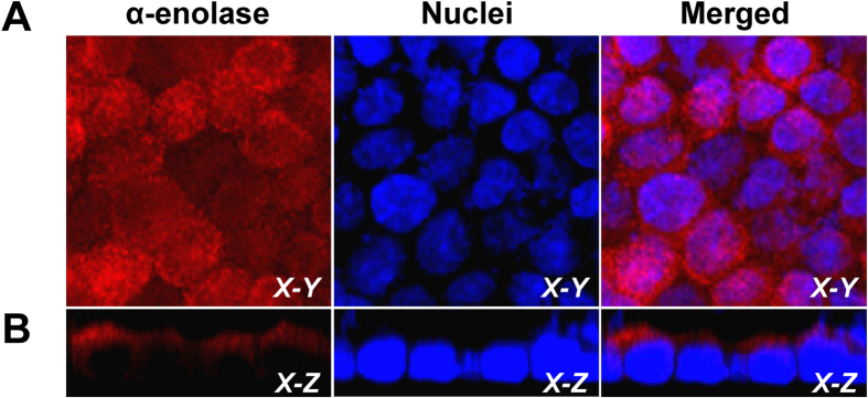
Figure 2. Confirmation of apical membrane localization of α-enolase on polarized MDCK cells.
The polarized MDCK cell monolayer was fixed with 3.7% formaldehyde (without permeabilization) and then incubated with rabbit polyclonal anti-α-enolase antibody followed by incubation with Cy3-conjugated anti-rabbit IgG secondary antibody containing 0.1μg/ml Hoechst dye for nuclear staining. The confocal micrographs were obtained from horizontal (X-Y) sections at apical membranes (A) and also sagittal (X-Z) sections (B). Original magnification power was 630X for all panels. Expression of α-enolase is shown in red, whereas nucleus is illustrated in blue.

