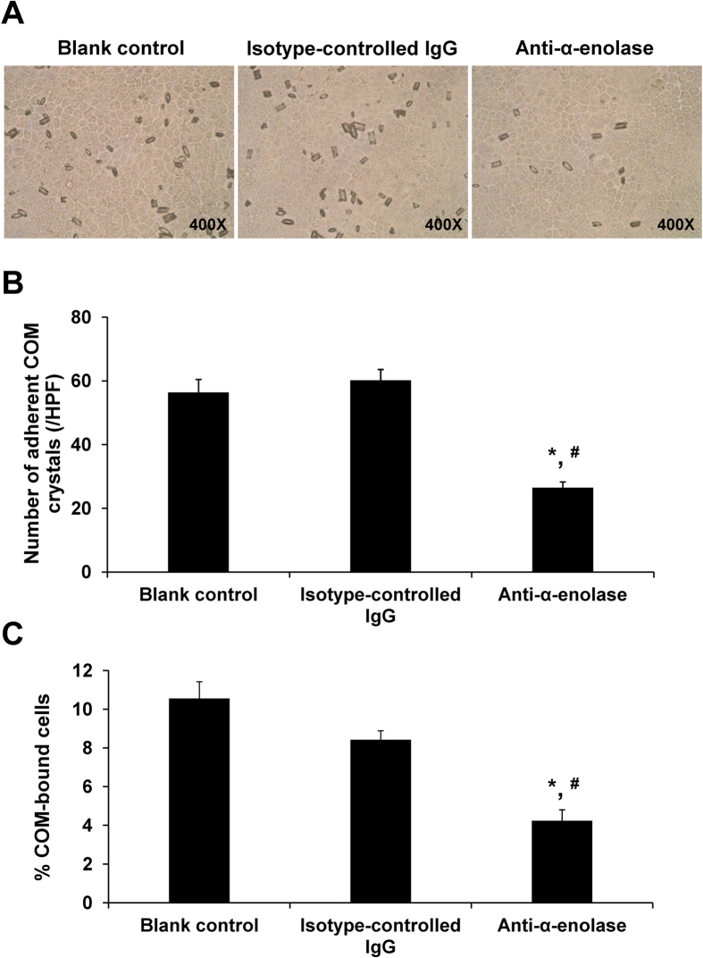
Figure 3. Cell-COM crystal adhesion assay and neutralization by a specific anti-α-enolase antibody.
The confluent polarized cell monolayer was incubated with 0.2 μg/ml anti-α-enolase antibody or 0.2 μg/ml rabbit isotype-controlled IgG prior to cell-crystal adhesion assay (see details in “Materials and Methods”), whereas the cells without antibody pretreatment served as the blank control. (A) After removal of unbound crystals, the adherent crystals remained on the cell surface were imaged by a phase-contrast microscope. (B) The adherent crystals were counted from at least 15 random high power fields (HPFs). (C) Percentage of COM-bound cells was also determined. Original magnification power was 400X. Each bar represents mean ± SEM of the data obtained from 3 independent experiments. *p < 0.05 vs. blank control; #p < 0.05 vs. isotype-controlled IgG.

