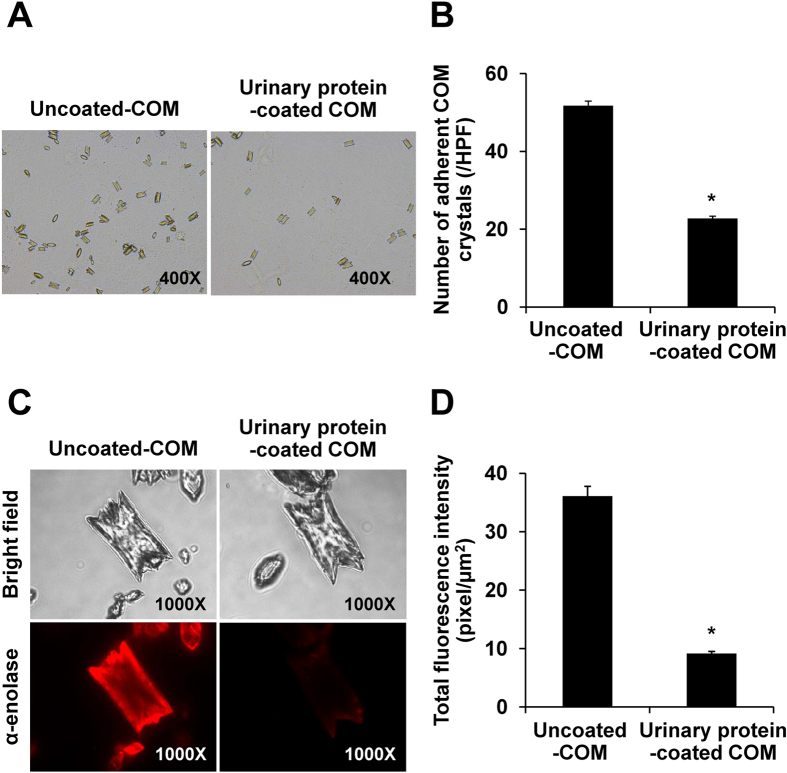
Figure 6. Effects of urinary proteins on cell-COM crystal adhesion and crystal-protein binding.
(A,B) Cell-COM crystal adhesion assay (original magnification = 400X). The adherent crystals were counted from at least 15 random high power fields (HPFs). (C,D) Crystal-protein binding assay (original magnification = 1,000X). Quantitative analysis of immunofluorescence staining of α-enolase bound on COM crystals in each group was performed using ImageJ software from at least 100 individual COM crystals per group. Each bar represents mean ± SEM of the data obtained from 3 independent experiments. *p < 0.05 vs. uncoated-COM crystal.

