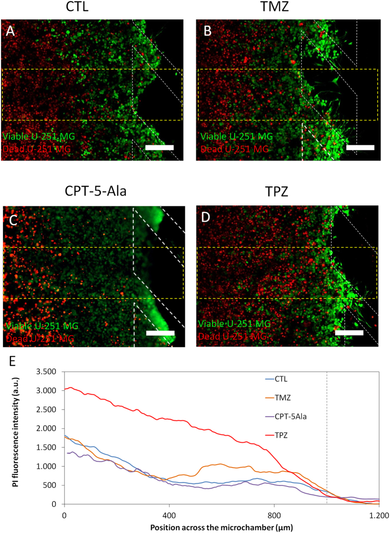
Figure 8. Drug effect on U-251 MG cells.
U-251 MG cells were cultured within the central microchamber for 24 hours before drug addition in order to allow time for formation of a hypoxic region first. Growth medium (A), 100 μM TMZ (B), 1.6 μg/ml CPT-5Ala or 100 μM TPZ (C) were perfused through both lateral microchannels and after 3 days cell viability was assessed using by PI/CAM staining. (E) The graphs show the PI fluorescence intensity along the delimited region, demonstrating that TMZ had a mild effect on U-251 MG cell viability. TPZ exerted a strong effect in the hypoxic areas. Position of the pillars is delimited by a grey dashed line. Scale bar is 200 μm.

