Fig. 4.
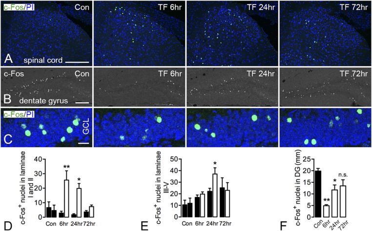
Modulation of c-Fos expression in the spinal cord and hippocampal formation after unilateral tibial fracture. (A) Robust activation of c-Fos in the superficial layers (laminae I and II) of the dorsal horn peaking at 6 h after fracture and declining to basal levels after 72 h. The activation of c-Fos in deep layers of the spinal cord appears increased bilaterally, but significance is reached only at 24 h compared with the contralateral side. (B and C) c-Fos expression in granule cells of the dentate gyrus is decreased transiently (6, 24 h) after tibial fracture, returning to basal levels after 72 h, as substantiated by quantification. Only the number of c-Fos–positive nuclei was counted, without considering intensity. Projection pictures in C are produced from 12- to 14-μm z-stack scanning with an interval of 2 μm. Con, control; GCL, granule cell layer; PI, propidium iodide; TF, tibial fracture. n = 14; n = 3 for the control group; n = 4 for both 6- and 24-h injury; and n = 3 for 72-h injury. Data are represented as mean ± SD. *P < 0.05, **P < 0.01; unpaired t test was applied for c-Fos in the spinal cord, and one-way ANOVA followed by Dunnett’s multiple comparison test was applied for c-Fos in the hippocampus with tibial fracture. [Scale bars, 200 μm (A and B) and 20 μm (C).]

