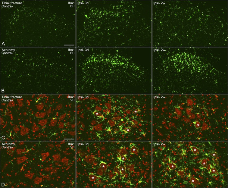
Fig. S4.
Activation of microglia in the spinal cord 3 d and 2 wk after unilateral injury. (A and B) Confocal micrographs show activation of microglia in the dorsal horn after fracture, with regard to both morphology and numbers (A) and more so after axotomy (B). (C and D) Projection micrographs show activated cells in the ventral horn, especially in the area surrounding motor neurons. After fracture, an increase is only seen after 3 d; but the activation of microglia is maintained even after 2 wk of axotomy. Counterstaining with propidium iodide (red) (C and D). Projection micrographs are produced from z-stack scanning of six series pictures with an interval of 2 μm. Asterisks indicate motor neurons surrounded by activated microglia. [Scale bars, 100 μm (A and B) and 50 μm (C and D).]

