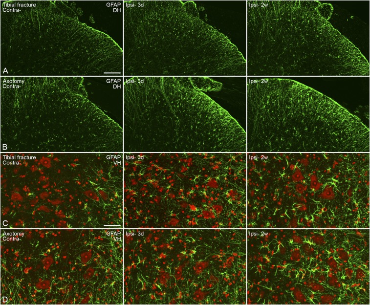
Fig. S5.
Activation of astrocytes in the spinal cord 3 d and 2 wk after unilateral tibial fracture or axotomy. (A and B) Confocal micrographs show modest activation of astrocytes in the dorsal horn, with regard to both morphology and numbers (A) and more so after axotomy (B). (C and D) Projection micrographs show activated cells in the ventral horn, especially in the area surrounding motor neurons (C), again stronger after axotomy (D). Counterstaining with propidium iodide (red) (C and D). Projection pictures are produced from z-stack scanning of six series pictures with an interval of 2 μm. [Scale bars, 100 μm (A and B) and 50 μm (C and D).]

