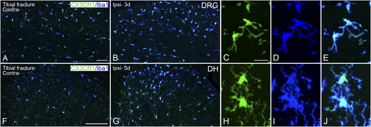
Fig. S6.
Increased expression of CX3CR1-LI in microglia in DRGs and spinal dorsal horn 3 d after unilateral tibial fracture. Confocal micrographs show double staining for Iba1-LI (blue) and CX3CR1 (green) in activated microglia in DRGs (A–E) and dorsal horn (F–J). High-magnification, projection micrographs show colocalization of CX3CR1/Iba1 in microglia from the ipsilateral ganglion (C–E) and in the ipsilateral dorsal horn (medial part) (H–J). Projection micrographs are produced from z-stack scanning of 13/14 series micrographs with an interval of 1 μm. [Scale bars, 200 μm (A and B), 500 μm (F and G), and 20 μm (C–E and H–J).]

