Fig. 3.
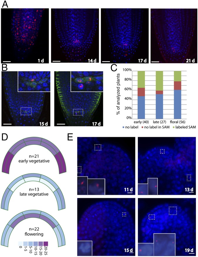
Retention of label-retaining cells within plant meristems. (A) EdU labeling time course. Roots were pulse-labeled with EdU for 1 h, 2 d after imbibition and transferred to plates lacking EdU. Cells that rapidly proliferated lost labeled chromosomes, whereas those that divided more slowly retained the label. Blue: DAPI; Red: EdU. (B) Colocalization of EdU-labeled nuclei with a WOX5:GFP (green) reporter that labels quiescent center (QC). The majority of the colocalized signals were located above the WOX5-labeled QC (Left), although QC cells were also labeled (Right). (C) Frequency of labeling of plants in the SAM. Plants were pulse-labeled with EdU for 1 h and then transferred to soil for 6–10 d (early vegetative), 11–14 d (late vegetative), or 13–19 d (flowering) at which time they were fixed, embedded in paraffin, and sectioned. Plants were scored as stained if EdU staining was visible anywhere in the aerial portion of the plant. (D) Heat map indicating the percentage of labeled plants at each stage with EdU-positive cells within the indicated zones of the SAM. (E) Examples of EdU-labeled SAMs with the days after the initial pulse indicated. (Top Left) Labeling within the L1 layer. (Bottom Left) Labeled cell in the L2 layer entering mitosis. (Top Right) Central section of a floral meristem with labeled cells in the L2 layer. (Bottom Right) Inflorescence meristem with several cells labeled in the L2 layer.

