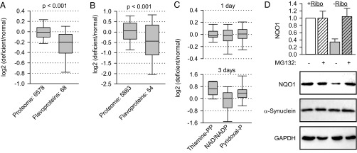
Fig. 1.
Depletion of riboflavin destabilizes the flavoproteome. (A and B) B16 murine melanoma cells were incubated in normal and riboflavin-deficient medium for 24 h (A) or 3 d (B). Then their proteomes were compared by quantitative mass spectrometry. Average changes from four experiments for each protein in the two groups are plotted. The significance of the difference between the medians of the two groups was assessed by Mann–Whitney test and is indicated. (C) Average changes in protein groups containing the indicated cofactors were determined after 1 d or 3 d. (D) Degradation of endogenous NQO1 in B16 cells upon riboflavin depletion (−Ribo) for 24 h (n = 3, mean ± SD). The stability of endogenous α-synuclein was analyzed. GAPDH was used as loading control.

