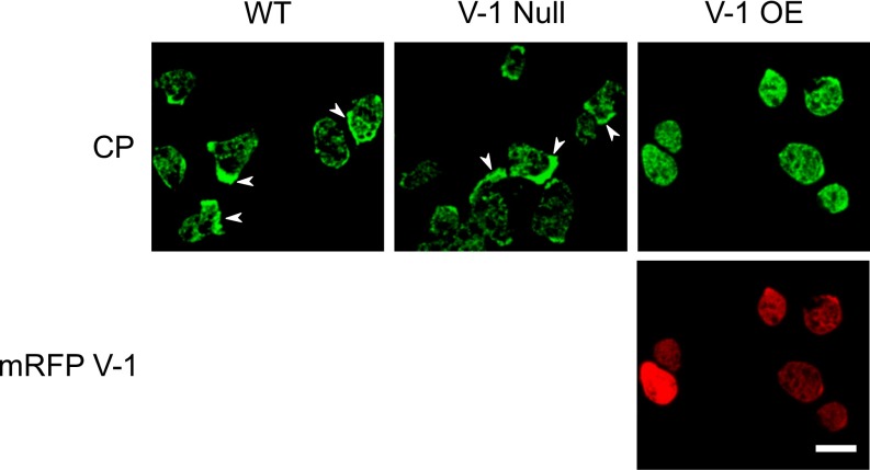
Fig. S5.
Localization of CP in WT, V-1–null, and mRFP-V-1–overexpressing cells. Shown are representative fields of WT, V-1–null, and mRFP-V-1–overexpressing cells stained with the antibody to CPα. Whereas WT and V-1–null cells show a preponderance of cortical localization for endogenous CP (arrowheads), cells overexpressing mRFP-V-1 show a preponderance of cytoplasmic localization for endogenous CP. (Scale bar, 10 µm.)

