
Figure 2. Whole-brain changes in functional connectivity to the right amygdala in bipolar mania compared to euthymia.
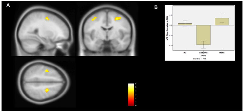
A The right amygdala demonstrates increased functional connectivity to the bilateral supplementary motor area (BA6) in bipolar mania compared to euthymia. This region of significantly increased connectivity is shown projected onto a MNI152 template brain at the x28, y-6, and z50 levels The color bar indicates T-statistic magnitude.
B A bar chart of the average Fisher’s Z transformed functional connectivity values between the right amygdala ROI and the combined BA6 clusters among the subjects within each group. Error bars represent standard errors.