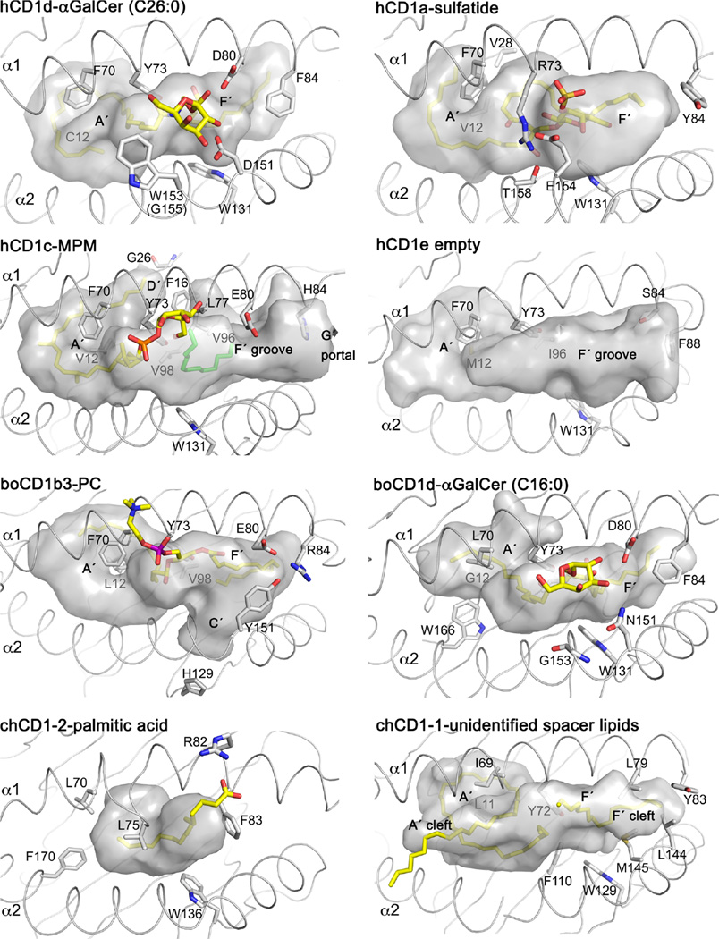
Fig. 3.
CD1-binding pockets and structural features. All CD1 proteins, expect for chCD1-2 have an A′ pole and at least the two pockets A′ and F′. Structural differences are the closed A′ roof of CD1a; the D′ portal, open F′ pocket, and G′ portal of CD1c; the open groove of CD1e; the T′ tunnel of CD1b; the partially closed A′ pocket of bovine CD1d; the lipid-binding pore of chCD1-2; and the A′ and F′ cleft of chCD1-1. CD1 residues beneath the binding groove are labeled in gray

