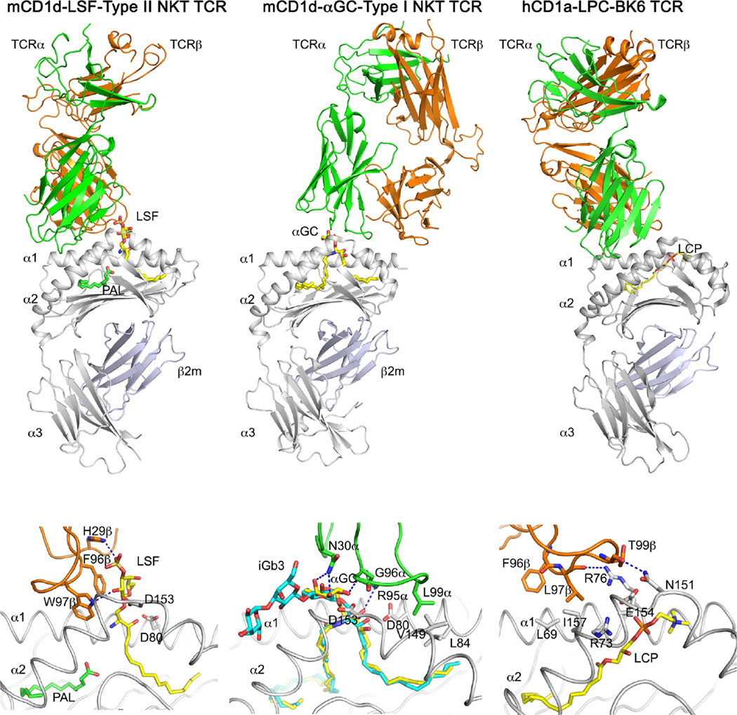
Fig. 4.
TCR recognition of CD1-presented antigens. Structures of the XV19.3 type II NKT TCR in complex with CD1d-lysosulfatide (LSF), the type I NKT TCR bound to mCD1d-presented αGalCer (αGC), and the BK6 TCR bound to CD1a presenting the permissive ligand lysophosphatidyl choline (LPC) are shown at the top, with detailed interactions shown below. Note that the BK6 TCR does not directly contact the lipid antigen, while both types I and II NKT TCRs require contacts with the antigen for binding. Hydrogen bonds in blue dashed lines; lipids in yellow and cyan; TCRα chain in green; TCRβ chain in orange; CD1 ingray. Note that the β-anomeric glucose of iGb3 is molded upon TCR binding into the approximate position of α-GalCer to allow for conserved TCR interactions (bottom middle panel)

