
Figure 3.
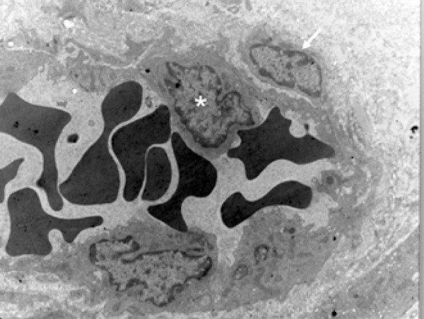
Electromicrography that illustrates the pericyte (arrow) in close contact with the endothelial cell (asterisk), sharing the basal membrane of this cell, which makes up the blood vessel wall (Scanning Electron Microscopy – 7,000X)
Official websites use .gov
A
.gov website belongs to an official
government organization in the United States.
Secure .gov websites use HTTPS
A lock (
) or https:// means you've safely
connected to the .gov website. Share sensitive
information only on official, secure websites.
Electromicrography that illustrates the pericyte (arrow) in close contact with the endothelial cell (asterisk), sharing the basal membrane of this cell, which makes up the blood vessel wall (Scanning Electron Microscopy – 7,000X)