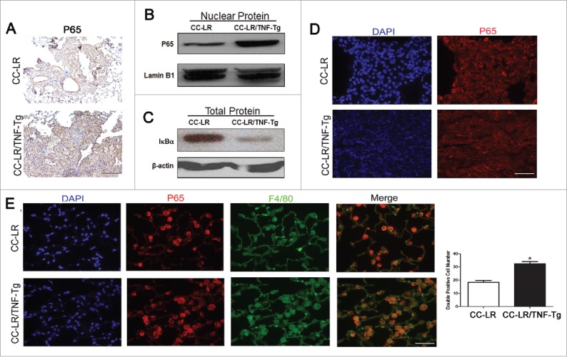
Figure 5.

TNF induces activation of NF-κB pathway in lung infiltrating macrophages and M-MDSCs. (A) Representative photomicrographs of P65 positive cells in lung tissue of CC-LR and CC-LR/TNF-Tg mice (20× magnification, scale bar = 50 μm, applicable to all panels). (B) Western blot analysis of P65 protein on the nuclear protein extracted from whole lung tissue of CC-LR and CC-LR/TNF-Tg mice. (C) Western blot analysis of IκBα on the total protein extracted from whole lung tissue of CC-LR and CC-LR/TNF-Tg mice. (D) Representative immunofluorescence staining showing P65 positive cells in lung tumors from CC-LR and CC-LR/TNF-Tg mice (40× magnification, scale bar = 25 μm, applicable to all panels). (E) Representative immunofluorescence photomicrograph (40× magnification, scale bar = 25 μm, applicable to all panels) and absolute number of P65 and F4/80 double positive cells in lungs of CC-LR and CC-LR/TNF-Tg mice (mean ± SE; *p < 0.05).
