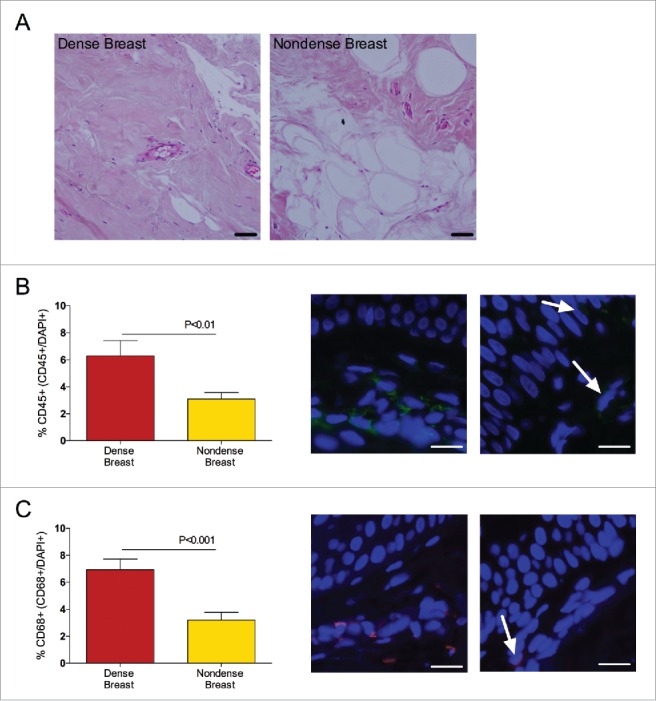
Figure 3.

Dense breast tissue contains increased levels of inflammatory cells in 39 postmenopausal women, attending the regular mammography-screening program who underwent biopsies for research purposes. On their regular screening mammograms, the breast were categorized as either dense or nondense and thereafter the women were invited to participate in the study as described in the materials and methods section. Breast biopsies were obtained in the upper lateral quadrant of the left breast. Tissue sections were stained and quantified as described in Materials and Methods. Graphed data are presented as mean ± SEM. (A) Representative H&E-stained tissue sections from dense and nondense breast tissue, respectively. Scale bars = 50 μm. (B) Sections were stained with CD45 (green). Significantly increased levels of leucocytes were detected in dense breast tissue. Representative tissue sections from each group are shown. Arrows indicate CD45+ cells. Scale bars =100 μm. (C) Sections were stained with CD68 (red). Significantly increased levels of leucocytes were detected in dense breast tissue. Representative tissue sections from each group are shown. Arrows indicate CD68+ cells. Scale bars =100 μm
