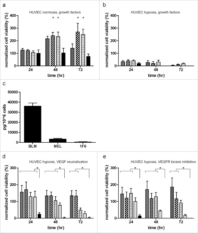
Figure 3.
Melanomas mediate endothelial cell (EC) survival under hypoxia, independent of the VEGF signaling pathway. (a,b) ECs were treated with 10 ng/mL VEGF ( ) or 200 ng/mL bFGF (
 ) alone, or in combination (
 ). Cell survival was monitored under normoxia (a) and hypoxia (b) for up to 72 h. (c) Levels of VEGF (pg/ 1 × 10⁶ cells) in serum-free melanoma conditioned medium (SF-CM) as detected by ELISA. Data were normalized to the number of cells used to generate the SF-CM and are expressed as mean ± SD of quadruplicate samples. (d, e) Effect of VEGF pathway inhibition on EC survival under hypoxia. ECs were treated with undiluted and twice diluted melanoma conditioned medium in the presence of (d) VEGF neutralizing antibody (0.3 µg/mL) or (e) VEGFR kinase inhibitor (0.3 µg/mL) (undiluted melanoma CM:
  ; undiluted melanoma CM with neutralizing antibody/inhibitor:
  ; twice diluted melanoma CM:
  ; twice diluted melanoma CM with neutralizing antibody/inhibitor:
  ; and basal medium:
  ). Normalized cell viability (%) data in (a, b, d, and e) is expressed relative to treatment with basal medium (DMEM + 10% FBS) at 24 h normoxia and represent mean ± SEM of three independent experiments, conducted in triplicate. *p < 0.05.

