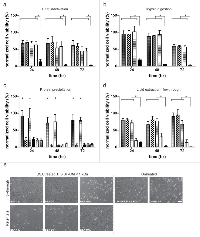
Figure 5.
Characterization of the melanoma-specific survival effect. (a) Effect of heat treatment on endothelial cell (EC) survival. Size fractionated< 1 kDa fraction of serum-free melanoma conditioned medium (SF-CM) was heat inactivated at 56°C, 80°C and 100°C, respectively, for 30 min. Untreated ( ), heat-inactivated (56°C:
  ; 80°C:
  ; 100 °C:
  ) media and basal medium (
 ) were added to near confluent EC cultures and long-term survival under hypoxia was monitored. (b) Effect of enzymatic digestion on EC survival. Size fractionated < 1 kDa fraction of melanoma SF-CM was treated with 100 and 200 µg/mL of trypsin for 30 min at 37 °C. Untreated (
 ), trypsin treated samples (100 µg/mL:
  ; 200 µg/mL:
  ) and basal medium (
 ) were added to near confluent EC cultures and monitored for long-term survival under hypoxia. (c) Effect of protein precipitation on EC survival. Size fractionated < 1 kDa fractions of melanoma SF-CM and basal medium were subjected to protein precipitation using acetone. Reconstituted protein and non-protein fractions were added to subconfluent EC cultures and survival under hypoxia was monitored (< 1 kDa SF-CM untreated:
  ; < 1 kDa SF-CM protein fraction:
  ; < 1 kDa SF-CM non-protein fraction:
  ; basal medium:
  ; basal medium protein fraction:
  ; basal medium non-protein fraction:
  ). (d,e) Effect of lipid extraction on EC survival. Size fractionated < 1 kDa SF-CM fractions of melanoma SF-CM were treated with 1%, 3%, or 10% fatty-acid free BSA. BSA-poor (flowthrough) fractions were obtained as described under ‘Materials and Methods’. (d) Untreated (
 ), BSA treated flowthroughs (1%:
  ; 3%:
; 3%:  ; 10%:
  ; ) and basal medium (
 ) were added to subconfluent ECs to monitor cell survival under hypoxia. (e) Morphology of ECs under various treatments at 48 h hypoxia. Scale bar, 100 µm. Normalized cell viability (%) in (a–d) is expressed relative to survival of basal medium (serum-free DMEM) treated ECs at 24 h normoxia and represent mean ± SEM of four independent experiments, conducted in triplicate. *p < 0.05.

