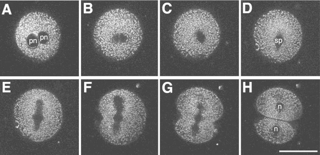
Figure 4.
Imaging the first cell division. MPLSM images of a Mitotracker-labeled pronucleate embryo undergoing the first cleavage division. The closely apposed pronuclei begin to break down (A—18.6 h PI) and lose their shape (B—19.1 h PI). Then a spindle begins to form (C—19.6 h PI) and exhibits a more distinct structure (D—20.25 h PI). The spindle elongates (E—20.4 h PI) and a cleavage furrow initiates (F—20.6 h PI). Finally, the cleavage furrow bisects the spindle (G—20.9 h PI) to form two daughter cells (H—21.0 h PI). Note: pronuclear accumulation of mitochondria was observed in this embryo prior to the time of the first image shown in this series. pn: pronucleus, sp: spindle, n: nucleus. Scale bar = 50 µm.

