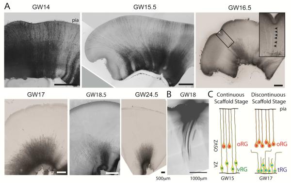
Figure 1. Morphological Transition in Radial Glia Scaffold During Human Cortical Development.
(A) DiI labeling of cells contacting the ventricular surface throughout neurogenesis reveals continuous radial glia scaffold prior to GW16.5, but only shorter “truncated” processes after GW16.5. Arrows indicate sporadic examples of individual fibers extending to the pial surface at GW16.5. See also Figure S1 and S2 for additional examples. (B) Conversely, DiI placement at the pial surface at GW18 labeled fibers extending to the OSVZ, but no ventricular surface contacting cells (See also Figures 3, S4). (C) Schematic representing three major classes of radial glia cells: classical ventricular radial glia (vRG) with fibers extending to the pial surface, ventricle contacting “truncated” radial glia (tRG) with non-classical morphologies whose processes terminate in the OSVZ, and outer radial glia (oRG).

