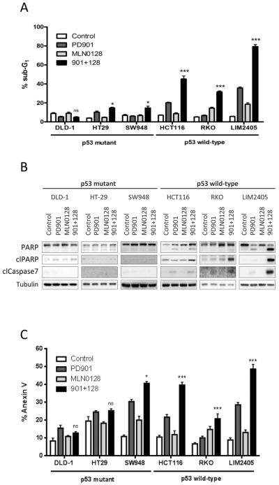
Figure 2. MEK- and mTOR-inhibition induces apoptosis in p53 wild-type CRC cells.
The indicated CRC cell lines were treated with DMSO (Control), 50 nM PD901, 50 nM MLN0128 or the combination of both inhibitors (901 + 128). A. Apoptosis was measured after 72 hours of treatment as the percentage of cells with sub-G1 DNA content by flow cytometry and analyzed with FCS Express 4 Flow software. Data are expressed as mean ± S.E from three independent experiments. B. Whole-cell protein extracts were analyzed after 24 hours of treatment by Western blotting with the indicated antibodies. Tubulin antibody was used as loading control. Figures are representative of three independent experiments. C. Apoptosis was measured after 72 hours of treatment quantification of the Annexin V positive cells (Guava Nexin Reagent, Millipore). n.s., not significant, *p<0.5, ***p<0.001.

