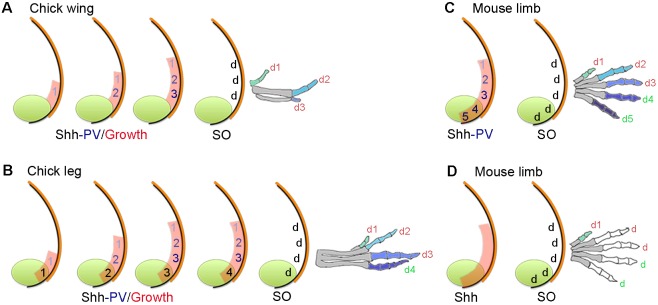
Fig. 1.
Positional information and self-organization in digit patterning. (A) In the chick wing, graded paracrine Shh signalling (numbers shaded blue) from the polarizing region (green) promotes growth of the digit-forming field (red), and in a positional information model, specifies cells with the three positional values (PV) 1, 2 and 3. Cells are specified with anterior positional values and promoted to posterior values every 4 h to give rise to three digits (d) by self-organization (SO). Note that limb buds are not drawn to scale. In all cases, colours on digits indicate a different positional value with which cells were specified, which are interpreted into phalange number (metacarpals are shaded grey). (B) In the chick leg, patterning is as in the chick wing (A) but a digit is derived from the polarizing region (green number), which is specified by the duration of non-graded autocrine Shh signalling (black numbers). (C,D) In the mouse limb, two digits are derived from the polarizing region (note digit 1 positional value is considered independent of Shh), positional values specified by Shh signalling (C) or not specified by Shh signalling (D) and self-organization produces four digits (2-5). Note, in C and D Shh signalling specifies the size of the digit-forming field (red shading).

