Fig. 7.
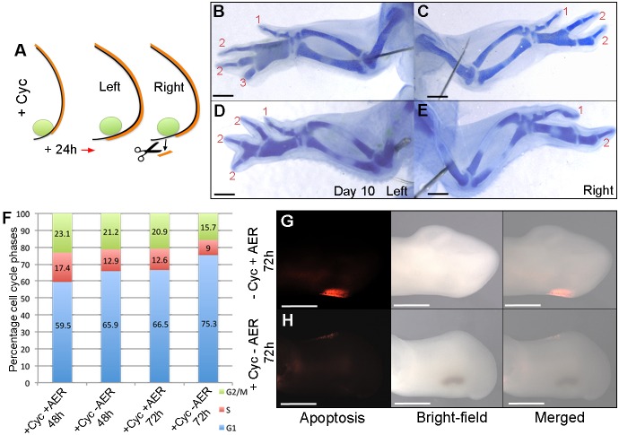
AER supports polarizing region digit formation. (A) Cyclopamine was applied at HH20/21 and posteriorly extended AER removed in right-hand wing buds after 24 h. 1-2-2 (C) or 1-2 (E) digit patterns develop. Note four digits in left wings of same embryos (B,D; n=15/15; see Table S5). Pearson's χ2 test reveals a significant increase in G1-phase cells in the polarizing region at 48 h (P<0.0001) and 72 h (P<0.0001) in HH20/21 cyclopamine-treated wing buds with the posterior AER removed compared with HH20/21 cyclopamine-treated wing buds with an intact posterior AER (F), indicating a reduced rate of proliferation. Apoptosis is detectable in the posterior necrotic zone of untreated wing buds (G, n=6/6) but undetectable in HH20/21 cyclopamine-treated wing buds in which the AER was removed (H, n=8/8). Scale bars: 1 mm (B-E), 500 μm (G,H).

