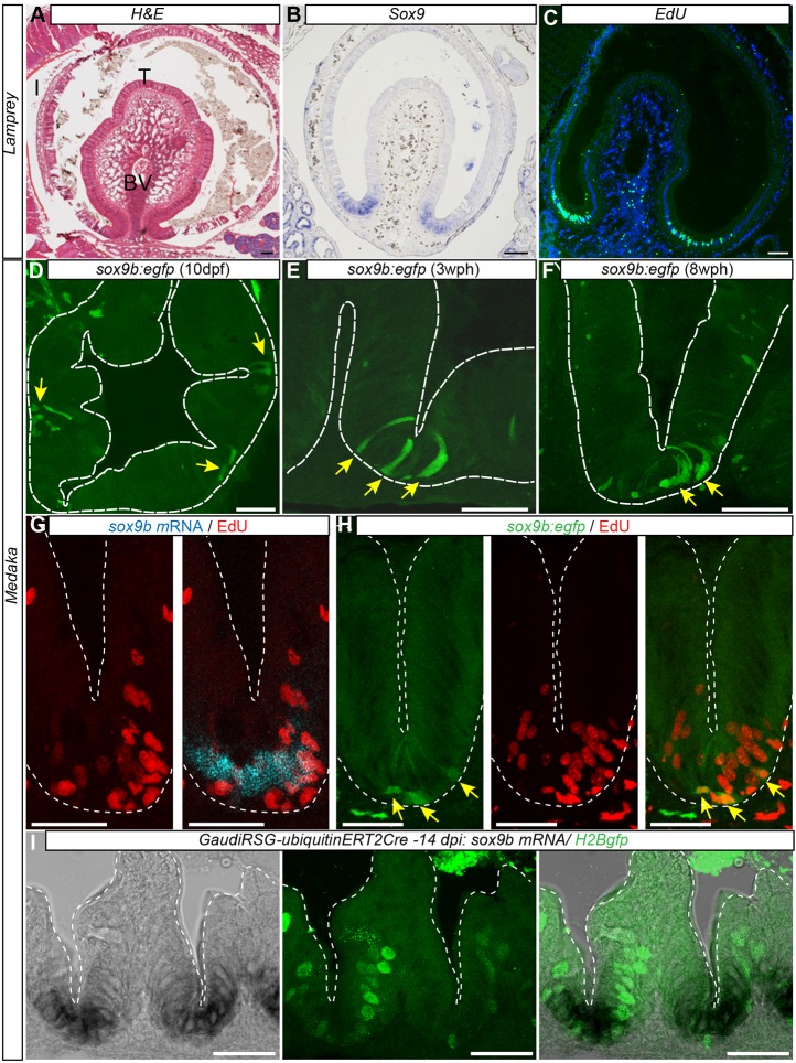
Fig. 3.
Expression pattern of intestinal sox9 is conserved from lamprey to medaka. (A) Transverse section of lamprey larval intestine stained with H&E, highlighting the typhlosole as a single fold. (B) In situ detection of Sox9 expression at the base of the typhlosole. (C) EdU+ cells (24 h after injection) reveal basal proliferation zone (green; nuclei: DAPI, blue). (D-F) Medaka Sox9b:eGFP-expressing cells mark proliferative intestinal cells at the base of furrows. Confocal images of transverse cryosections of sox9b:gfp transgenic intestine at 10 dpf (D) in 3-week-old juveniles (E) and 8-week-old adult fish (F). Arrows indicate position of sox9b:eGFP-expressing cells at the base of folds. (G) Colocalization of endogenous sox9b expression domain shown by in situ hybridization and proliferation in EdU+ cells. (H) Colocalization of sox9b:eGFP-expressing cells and EdU staining. Arrows indicate sox9b:eGFP-expressing cells. Note that sox9b:eGFP-expressing cells are EdU+. (I) Sox9b endogenous expression shown by in situ hybridization in GaudíRSG-ubiquitin:ERT2Cre transgenic fish (left panel), clonal analysis of GaudíRSG-ubiquitin:ERT2Cre transgenic fish 2 weeks after induction. At the base of the furrow, proliferating GFP+ stem cells (middle panel) express sox9b (right panel). Scale bars: 100 µm (A-C); 25 µm (D,G,H,I); 50 µm (E,F). BV, blood vessel; I, intestine; T, typhlosole.

