Fig. 4.
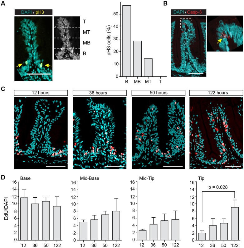
Intestinal stem cells are located in furrows between intestinal folds. (A) pH3 immunostaining on transverse sections of fold. Fold was divided into four equally sized regions: base (B), mid-base (MB), mid-tip (MT) and tip (T). Arrows in left panel indicate pH3+ cells. Respective frequency of pH3+ cells is shown in right panel. The majority of pH3+ cells are seen at the base of the fold. (B) Caspase-3 immunostaining on transverse sections of a fold. Caspase-3+ cells located at fold tip. (C,D) EdU pulse-chase assay. Adult fish were incubated in EdU for 24 h and fixed after 12, 36, 50, 122 h. For each time point, a representative fold from anterior and mid gut is shown in C. (D) Frequency of EdU+ cells in each region was analysed with KNIME. For each time point, three fish were analysed and nuclei from around 50 folds were counted for each region. Values are mean±s.e.m. A significant difference between 122 and 12 hours in the tip region was found using unpaired Student's t-test; P=0.028. Scale bars: 50 µm.

