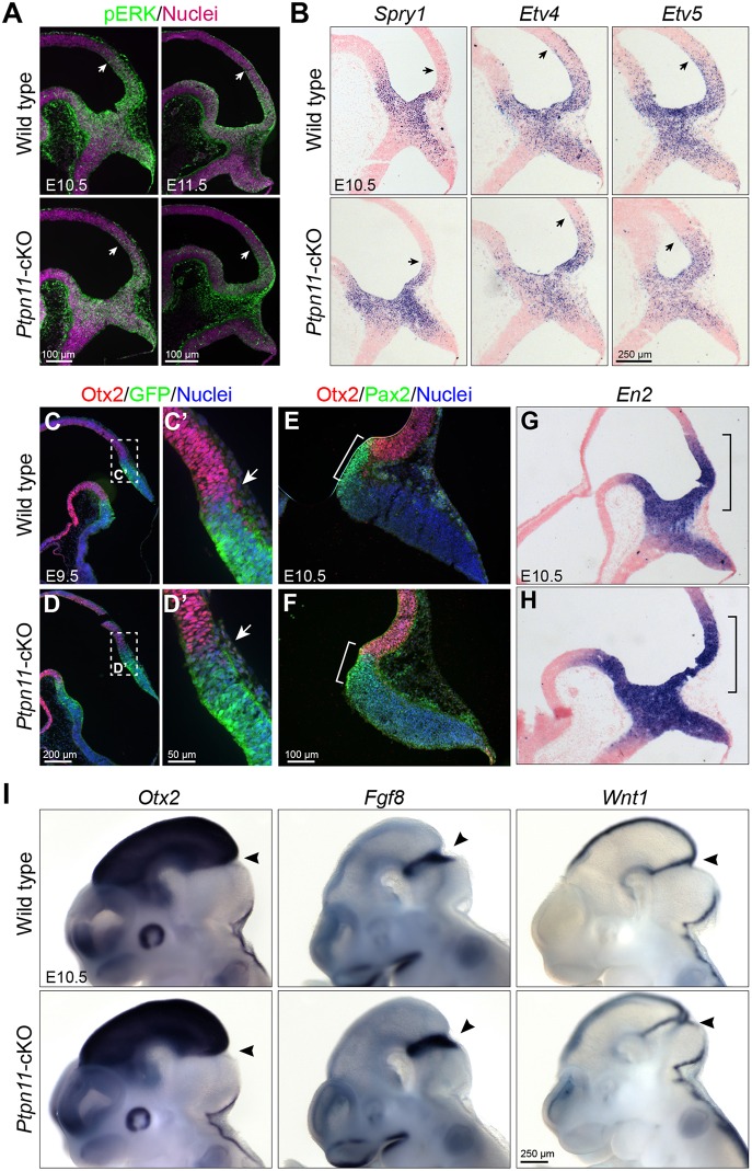
Fig. 2.
Pattern formation of the mes-r1 area is normal in Ptpn11-cKO embryos. (A) Immunofluorescence for pERK on sagittal sections. (B) In situ hybridization for Spry1, Etv4 and Etv5. Arrows indicate the anterior limit of detected signals in A,B. (C-F) Immunofluorescence on sections of E9.5 embryos carrying the Gbx2creER allele (C-D′) and E10.5 embryos (E,F). The boxed areas are enlarged in C′ and D′; arrows indicate the border between Otx2+ and GFP+ cells. The brackets demarcate the Pax2 expression domain. (G-I) In situ hybridization on sections (G,H) and whole mount (I) of E10.5 embryos. Arrowheads indicate the isthmus; brackets demarcate the En2 expression domain in the posterior mesencephalon.

