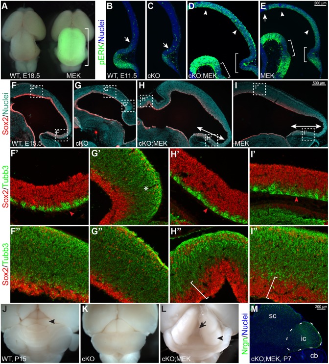
Fig. 5.
Expressing Mek1DD rescues the tectal stem zone and inferior colliculus in Ptpn11-cKO mutants. (A) Dorsal view of wild-type (WT) and En1cre/+;R26Mek1DD/+ (MEK) brains at E18.5. The bracket shows the enlarged tectum with GFP fluorescence, which marks Mek1DD expression. (B-E) Immunofluorescence for phosphorylated ERK (pERK) on sagittal sections of WT, Ptpn11-cKO (cKO), Ptpn11-cKO;MEK (cKO;MEK) and MEK embryos at E11.5. Arrows demarcate the anterior limit of pERK immunoreactivity; arrowheads show the pERK immunoreactivity throughout the mes; brackets show reduced intensity of pERK near the isthmus. (F-I″) Immunofluorescence on sagittal sections of E15.5 brains. The boxed areas in F-I are enlarged in F′-I″. The double arrows show the enlarged tectal stem zone (TSZ) with Mek1DD expression; the asterisk indicates increased accumulation of Tubb3+ cells; the arrowheads show the thin layer of Tubb3+ cells in the presumptive TSZ; the brackets denote the thickened ventricular zone. (J-L) Dorsal view of P15 brains. The arrowheads indicate the inferior colliculus (ic); the arrow shows the splitting of the tectum, a phenotype observed in mice with Mek1DD expression after birth. (M) Immunofluorescence on sagittal sections of P7 Ptpn11-cKO;MEK embryos. The IC is demarcated with a dashed line. cb, cerebellum; sc, superior colliculus.

