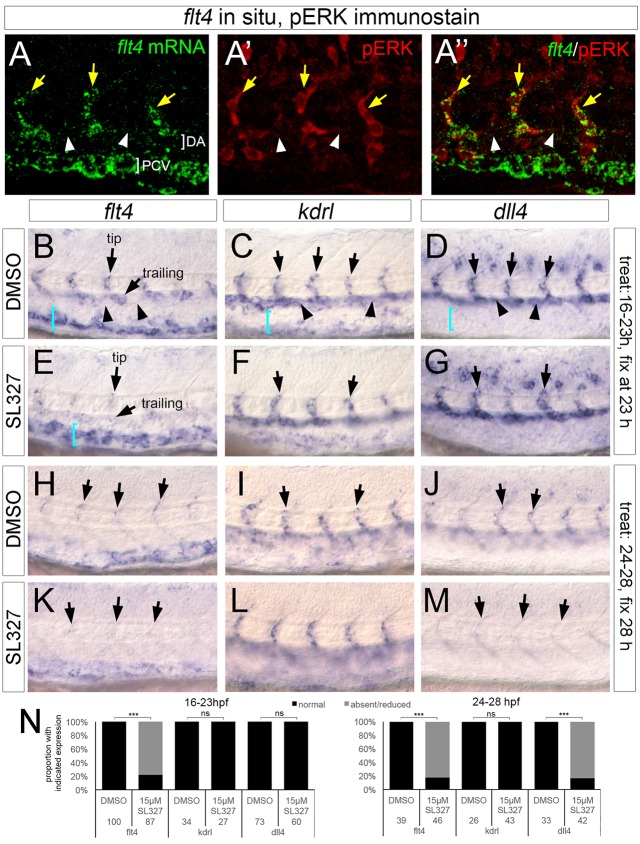
Fig. 5.
ERK activation is essential for flt4 and dll4 expression in sprouting ISVs. (A-A″) Confocal images of embryo co-stained for flt4 transcript (green) and pERK (red). Yellow arrows denote ISVs, white arrowheads indicate non-expressing cells in the dorsal aorta. (B-M) Whole-mount in situ hybridization with riboprobes against the indicated transcripts (flt4, kdrl or dll4). Unless otherwise noted, arrows indicate ISVs in all panels; in B and E, tip and trailing positions are noted by arrows. In B-D, arrowheads denote expression (or lack thereof) in intervening DA endothelial cells. In B-E, blue brackets indicate PCV. Compound used for treatment indicated at left, interval of treatment and analysis stage indicated at right. (N) Quantification of marker expression; treatment stages and concentrations are indicated, as are numbers of embryos used for analysis. ***P<0.001; ns, not statistically significant.

