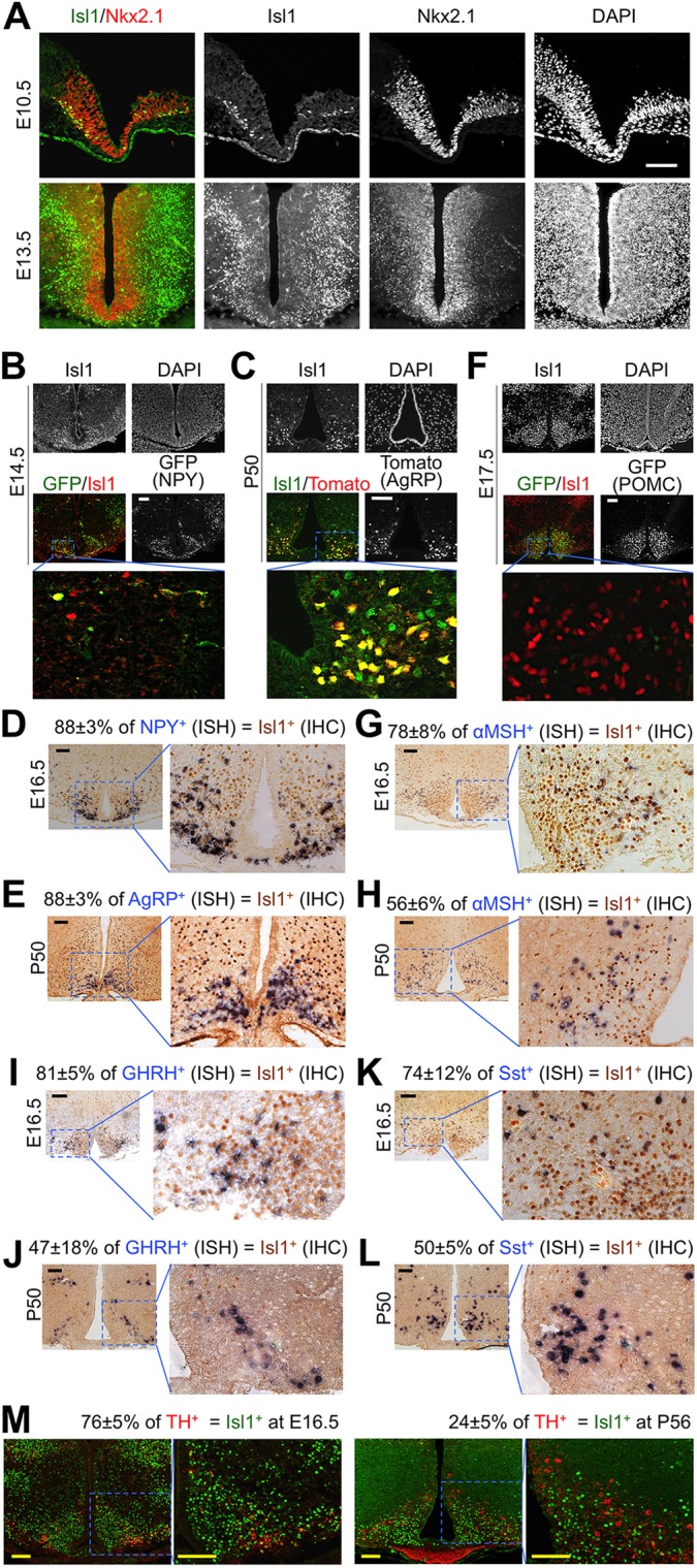
Fig. 1.

Expression of Isl1 in arcuate neurons. (A) Wild-type mouse embryonic hypothalamus was immunostained with anti-Isl1 and anti-Nkx2.1 antibodies. DAPI staining marks all nuclei in the section. (B,C,F) Representative images of the ARC from E14.5 Npy-hrGFP embryos (B), P50 Agrp-ires-Cre;Rosa26CAG-tdTomato mice (C) and E17.5 Pomc-eGFP embryos (F), which were immunostained with anti-Isl1 antibody. (D,E,G-L) ISH (blue) for the genes encoding NPY (D), AgRP (E), αMSH (G,H), GHRH (I,J) and Sst (K,L) was performed on either E16.5 or P50 wild-type mouse ARC, followed by immunostaining with anti-Isl1 antibody (brown). (M) Co-immunostaining of E16.5 and P56 wild-type mouse ARC with antibodies against TH and Isl1. Scale bars: 100 μm. (D,E,G-M) Values indicate the percentage of cells expressing the given marker that are also Isl1 positive (mean±s.d. of three hypothalami per panel).
