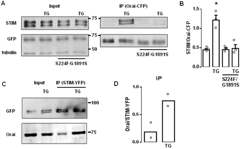
Fig. 2.
Mutant IP3Rs attenuate association of STIM and Orai after store depletion. (A) Western blots from brains of larval Drosophila expressing Orai–CFP with WT or mutant IP3Rs, and treated with thapsigargin (TG, 10 µM in Ca2+-free HBM for 10 min) as indicated. The input lysates (equivalent to 20% of the immunoprecipitated sample) and anti-GFP immunoprecipitates (IP) are shown. α-tubulin provides a loading control. The positions of molecular mass markers (kDa) are shown between blots. (B) Summary results for the ratio of the intensities of the STIM to Orai–CFP bands (mean±s.e.m., n=3). *P<0.05, paired Student's t-test relative to the respective control. (C) WT brains expressing STIM–YFP show results of immunoprecipitation (with anti-GFP antibody) after treatment with thapsigargin as indicated. The lysate lanes contain the equivalent of 20% of the immunoprecipitated sample lanes. (D) Summary results show the ratio of the intensities of the Orai to STIM–YFP bands (mean and individual values are shown; n=2).

