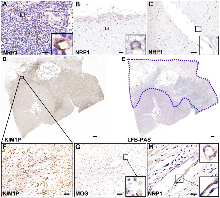
Fig. 5.
Endothelial NRP1 is induced in human acute multiple sclerosis lesions. (A) Immunohistochemistry analysis of NRP1 in normal human tonsil tissues reveals strong NRP1 expression in the endothelium. Images are representative of three technical replicates. (B) Normal human spinal cord samples showed limited NRP1 immunoreactivity in subpial regions. Enlarged view (inset) shows NRP1+ glial cells. (C) Normal human brain white matter shows no NRP1 signal. The enlarged view indicates NRP1− endothelial cells. (D) The low-magnification image of KIM1P immunohistochemical staining indicates extensive macrophage infiltration in white matter of a multiple sclerosis brain. (E) Luxol-Fast-Blue and periodic-acid–Schiff (LFB-PAS) staining on the multiple sclerosis tissue section reveals a destructive demyelinating lesion in the white matter (shown by the blue-dotted line). (F) The enlarged view of panel D shows a region of interest with numerous macrophage infiltrations (KIM1P staining). (G) Staining of myelin oligodendrocyte glycoprotein (MOG) in the same region as that shown in F indicates early active demyelinating activity, characterized by myelin debris with macrophages (enlarged view in the inset). (H) Increased NRP1 immunoreactivity in the reactive astrocytes and endothelial cells in the lesion shown in D–G (NRP1 staining). Results are representative of three samples in both normal human brains and multiple sclerosis lesions (B–H). Enlarged views in H show the positive staining of NRP1 in endothelial cells. Scale bars: 20 µm (A,B,H), 50 µm (C,F,G), 1 mm (D,E).

