Fig. 1.
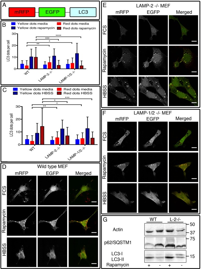
The autophagic flux is impaired in LAMP-2-single- and LAMP-1/2-double-deficient cells after autophagy induction. Cells were transfected with the tandem-fluorescent tagged mRFP–GFP–LC3 construct (A) and cultured in media (control), rapamycin (50 µm) (B) or HBSS (C) for 6 h. In wild-type MEFs, autophagy induction was characterized by an increased number of autophagosomes and autolysosomes (D) while in LAMP-2-single-deficient (E) and LAMP-1/2-double-deficient (F) cells the number of autolysosomes remained unchanged after treatment with rapamycin or HBSS. (G) Western blot analysis of total cell extract reveals an absence of degradation of p62 after rapamycin treatment confirming the blockage of the autophagic flux in LAMP-2-negative cells. Scale bars=10 µm. Data are expressed as mean±s.d. of three independent experiments. *P<0.05; **P<0.01; ***P<0.001; ****P<0.0001 (Mann–Whitney test).

