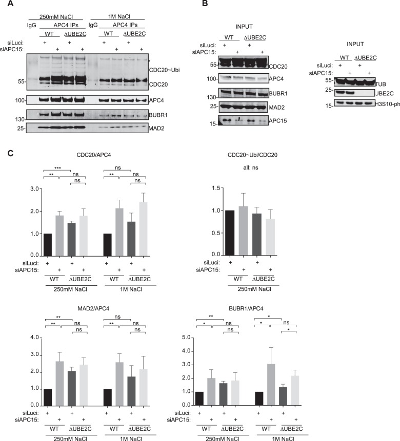
Fig. 4.
APC/C-MCC interaction in ΔUBE2C cells depleted of APC15. (A) The APC/C was purified in the presence of 250 mM NaCl or 1 M NaCl using an anti-APC4 antibody and purifications analyzed for MCC components by western blot. The asterisk indicates an unspecific band in Cdc20 blot. (B) Input for purifications in A. (C) Quantification of the levels of Cdc20, Mad2 and BubR1 normalized to APC4 and WT set to 1. Cdc20 ubiquitinated species are normalized to Cdc20. The mean±standard deviation (s.d.) is indicated. ns, non-significant; *P≤0.1; **P≤0.01; ***P ≤0.001 by t-test.

