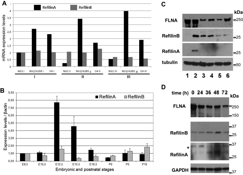
Fig. 3.
Differential regulation of RefilinA and RefilinB mRNAs and proteins. (A) Comparison of RefilinA and RefilinB mRNA levels in three cell sorting experiments (see Fig. S1C). (B) Comparison of RefilinA and RefilinB mRNA levels during mouse brain development. 1 µg of cDNA from embryonic and postnatal mouse brain [E8.5; E10.5; E12.5; E15.5; E18.5; postnatal day (P)0; P5; P10] were used to perform quantitative PCR. β-actin gene was selected as stable reference gene in embryo for internal standardization (Willems et al., 2006). Error bars represent the standard deviation (s.d.) of three independent experiments. We assigned the enrichment value of 1 for stage E8.5. (C) Western blot analyses of total cellular extracts of U373MG cells (lane 1), U373MG cells transfected with RefilinB or RefilinA constructs (lane 2), long term rat OPC cultures (lanes 3,5) or rat neural progenitors (lanes 4,6). OPC and neural progenitors were grown in Neurobasal medium supplemented with bFGF and PDGF (lanes 3,4) or bFGF and EGF (lanes 5,6). Anti-FLNA, anti-RefilinB, anti-RefilinA and anti-β-Tubulin antibodies were used as indicated. (D) Rat OPC cultures were shifted to differentiation culture medium for different period of time as indicated and lysed in SDS-sampling buffer. Total cell extracts (20 µg) were analysed by western blot using anti-FLNA, anti-RefilinB, and anti-GAPDH antibodies. A fivefold greater concentration of loaded sample was required to detect RefilinA (100 µg) with anti-RefilinA antibody.

