Fig. 5.

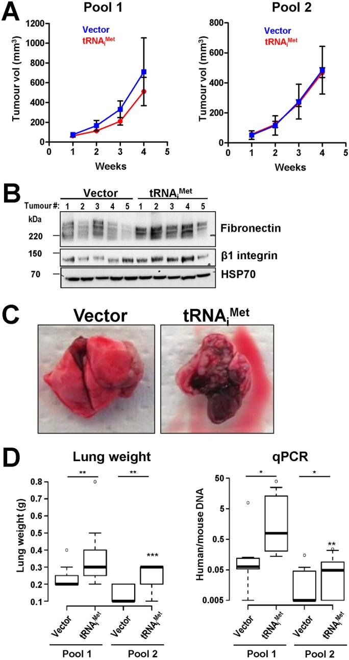
tRNAiMet drives melanoma metastasis, but not primary tumour growth. (A) WM266.4 cells stably expressing a vector encoding tRNAiMet (two independent pools) or an empty vector (Vector) (two independent pools) and were injected subcutaneously into the flank of CD1 nude mice. Subcutaneous tumour growth was measured by callipers three times a week and tumour volume was calculated from these. Values are mean±s.e.m. (B) tRNAiMet and empty vector expressing WM266.4 cells were injected subcutaneously into CD1 nude mice. The resulting tumours (5 from each condition) were lysed and their fibronectin and β1 integrin content determined by western blotting. HSP70 is used as a loading control. (C,D) tRNAiMet and empty vector expressing WM266.4 cells (two independent pools of each as for A) were injected via the tail vein into CD1 nude mice. The lungs of these animals were assessed for the presence of tumours by visual inspection (C), by determination of lung weight (n=7+7 pool 1 and 7+9 pool 2, pLHCX and tRNAiMet respectively) (D, left panel), and by qPCR to quantify the proportion of human genomic DNA (from the WM266.4 cells) with respect to mouse genomic DNA (from the host animal) (D, right panel). n=7+7 pool 1 and 7+11 pool 2, vector and tRNAiMet respectively. Values are expressed as box and whisker plots (whiskers: 5-95 percentile). The ratios of human to mouse DNA are expressed on a Log10 scale. **P<0.01; P<0.05; van Elteren Test (stratified Mann–Whitney). N.B. The injection of cells from pool 1 and pool 2 were conducted in experiments that were independent from one another.
