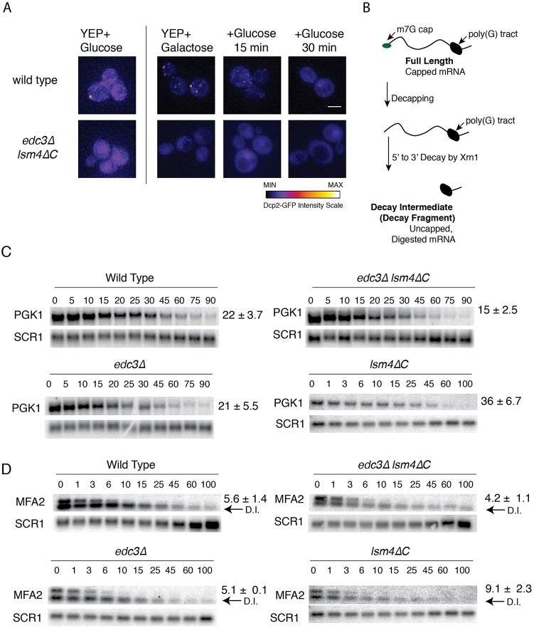
Fig. 1.
PGK1 and MFA2 mRNA stability in the edc3Δ lsm4ΔC mutant. (A) Wild type and edc3Δ lsm4ΔC mutant yeast cells expressing Dcp2-GFP from its endogenous locus grown in SC+2% glucose or 2% galactose as indicated. Later time points depict the cells grown in galactose after being washed, resuspended, and grown in medium containing glucose. (B) Depiction of the full length capped mRNA and the decay fragment generated by decapping and 5′ to 3′ degradation, which was ultimately blocked by a poly(G) tract inserted into the 3′ UTR. (C) Northern blots for the half-life determination of PGK1 mRNA in strains indicated. Time points (min) after transcriptional shut-off by glucose addition are shown. SCR1 is the loading control. Error=s.d., n=3-15 biological replicates. (D) As above for MFA2 mRNA, the half-life indicated to the right of the blots. Arrows labeled D.I. indicate the mRNA decay intermediate generated by decapping and 5′-to-3′ degradation blocked by the poly(G) tract in the 3′ UTR. Time points (min) are shown above the respective northern blots.

