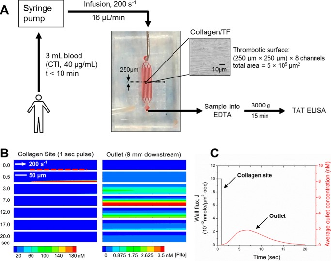
FIGURE 1.
Microfluidic setup for measuring thrombin flux during whole blood thrombosis on collagen/TF surface. A, a microfluidic device with 8 channels (each channel: 250-μm in width and 60-μm in height) diverging from a single inlet and converging into a single outlet was used for whole blood perfusion over collagen/TF surface. Blood was collected into CTI (40 μg/ml) and loaded into syringes that were subsequently mounted on a syringe pump. Blood infusion was initiated within 10 min of phlebotomy at a constant flow rate of 16 μl/min (initial wall shear rate = 200 s−1). Device outlet flow was collected into EDTA to quench thrombin generation with EDTA and allow for TAT formation. Blood samples (collected every 120 s) were subsequently centrifuged and analyzed by TAT ELISA. The microfluidic system was characterized with a COMSOL convection-diffusion model (B). To calculate the device response time, thrombin flux signal from a 250-μm-long surface domain with amplitude of 10−11 nmol/μm2-s was imposed as a boundary condition for duration of 1 s. Location of the surface domain is indicated by a red dashed line. Average thrombin concentration at the outlet peaked after a delay of 7 s and leveled off to zero by 20 s (C).

