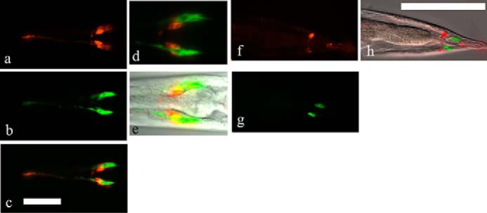
FIGURE 4.

Expression of the C41C4.1 gene product in vivo. EGFP signals (green) were distributed in amphid sheath cells in the head region (b–e) and in phasmid sheath cells in the tail region (g and h) of transgenic worms. DiI (red) was taken in by neuronal cells (amphid/phasmid neurons) near the sheath cells. a, amphid neurons stained with DiI. b, amphid sheath cells in the head region expressing C41C4.1::egfp fluoresce brightly. c, merge, the distribution of the two signals was generally distinct. d, another merged photograph showing that the C41C4.1 gene is expressed in amphid sheath cells (green) adjacent to amphid neurons (red). e, image overlaid with the corresponding DIC image. f, in the tail region of transgenic worms, phasmid neurons (red) were stained with DiI. g, cells expressing C41C4.1::EGFP are found near the phasmid neurons in f. h, DIC image overlaid with f and g. Magnification: a–c, ×400; d–h, ×1000. Bar: 50 μm.
